

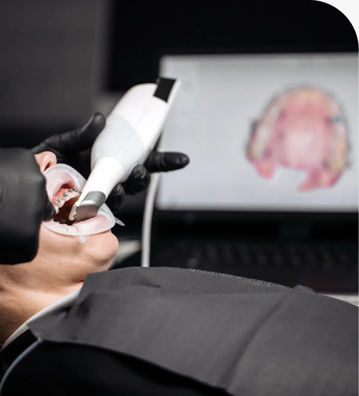
At Star City Family Dentistry, we use an intraoral camera to help patients see exactly what our dentist and team see. This small, handheld device captures high-resolution images inside the mouth and displays them on a screen in real time. Patients gain a clearer understanding of their oral health, treatment needs, and home care priorities without guesswork.
Intraoral Camera Explained
An intraoral camera is a pen-sized, chairside dental camera with LED lighting and a macro lens. It takes magnified photos and live video of teeth, gums, and other oral structures. Unlike a traditional mirror, it shows fine details such as early cavities, cracked enamel, worn fillings, plaque and tartar, and gum inflammation. Images can be paused, saved to your digital dental record, and compared over time to track changes.
Because it is a diagnostic dental technology, the intraoral camera supports more accurate diagnosis and planning. It also serves as a patient education tool. When you can view your own tooth surfaces at high magnification, recommendations for a filling, crown, or periodontal care become easier to understand.
Benefits of Intraoral Camera Imaging
- High-resolution images that reveal early problems before they worsen.
- Real-time viewing that encourages informed decisions and questions.
- Magnification that helps identify cracks, leakage around fillings, and calculus.
- Documentation that aids treatment planning and follow-up comparisons.
- Comfortable, noninvasive imaging with no radiation exposure.
- Helpful visuals for insurance claims and referrals when needed.
How Intraoral Camera Works
During an exam or cleaning, our dentist or hygienist places a protective barrier over the camera tip and gently positions it to view different areas of your mouth. The live image appears on a nearby monitor. When something important is seen—such as a chip, stain, cavity, or irritated gum tissue—a still photo is captured for your record. The device uses light and optics, not X-rays, so it does not replace radiographs; instead, it complements them by showing surface detail that X-rays may not reveal.
If you are curious about how an intraoral camera works moment to moment, think of it as a tiny high-definition video camera designed for close-up images. It is quiet, quick, and easy to reposition so multiple angles can be reviewed in seconds.
What to Expect During Your Visit
Expect a comfortable, quick process. You may feel light cheek retraction or gentle movement as the camera is positioned, but there is no pain. Images are displayed instantly so you can discuss what is shown and ask questions right away. The team will save photos to your chart to monitor changes, demonstrate home care techniques, or explain options if treatment is recommended. Most imaging takes only a few minutes and fits smoothly into a routine checkup.
Patients often appreciate seeing before-and-after images—for example, a stained groove before a sealant is placed or a fractured cusp prior to a crown. Clear visuals support confident decisions and help set expectations for care.
When Is It Used?
The intraoral camera is helpful during comprehensive exams, emergency visits for a chipped or painful tooth, assessments of gum health, and post-treatment checks. It assists with early cavity detection, evaluation of crown and bridge margins, documentation of wear from clenching or grinding, and confirmation of plaque buildup in hard-to-see areas. If you have specific concerns, you can request images of those areas for review.
Frequently Asked Questions About Intraoral Camera
Schedule Your Appointment
Welcoming New Patients
Need a checkup or treatment? Our team is ready to help you feel comfortable, informed, and cared for today.
Contact our Roanoke dentist and team today to reserve your visit and experience patient-focused care designed around your needs.